Introduction
Increasing interest in – and rapid development of – a wide range of materials and products containing nanoscale structures and engineered nanoparticles has been accompanied by a greater awareness that the longer term potential toxic effects of such materials and their potential environmental impact are poorly understood. Existing methods have been assessed and new methods sought by which such materials could be analyzed on a routine basis during development and manufacture. The use of Nanoparticle Tracking Analysis as a rapid and information-rich multi-parameter nanoparticle characterization technique which allows the user to obtain number frequency particle size distributions of polydisperse nanoparticulate systems has resulted in its rapid adoption as an interesting new technique in a wide range of sectors within environmental and toxicity studies. This white paper addresses some of the latest work in the literature in which NTA has been proposed, used and assessed in the study of nanoparticle toxicity and environmental impact.NTA has found use in a variety of investigations researching the toxicity and environmental impact of nanoparticles. As well as being used to determine the size of particles in investigations into the toxicity of carbon nanotubes and nanoparticulate metals, NTA has also been used in investigations on the interactions of nanoparticles with organisms at a cellular level and the development of methods for the testing of toxicity. NTA has proved to be a useful tool in determining both particle size and concentration of nanomaterials in waste water analysis.
Nanoparticle Tracking Analysis (NTA) Overview
NTA utilizes the properties of both light scattering and Brownian motion in order to obtain the particle size distribution of samples in liquid suspension. A laser beam is passed through the sample chamber, and the particles in suspension in the path of this beam scatter light in such a manner that they can easily be visualized via a 20x magnification microscope onto which is mounted a camera. The camera, which operates at approximately 30 frames per second (fps), captures a video file of the particles moving under Brownian motion within the field of view of approximately 100 μm x 80 μm x 10 μm.
The movement of the particles is captured on a frame-by-frame basis. The proprietary NTA software simultaneously identifies and tracks the center of each of the observed particles, and determines the average distance moved by each particle in the x and y planes. This value allows the particle diffusion coefficient (Dt) to be determined from which, if the sample temperature T and solvent viscosity η are known, the sphere-equivalent hydrodynamic diameter, d, of the particles can be identified using the Stokes-Einstein equation (Equation 1).
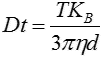
where KB is Boltzmann’s constant. NTA is not an ensemble technique interrogating a very large number of particles, but rather each particle is sized individually, irrespective of the others. An example of the size distribution profile generated by NTA is shown in Figure 2.
Figure 2: An example of the size distribution profile generated by NTA. The modal size for this sample is found to be approximately 70 nm, with larger sized particles also present. In addition, the particles’ movement is measured within a fixed field of view (approximately 100 μm by 80 μm) illuminated by a beam approximately 10 μm in depth. These figures allow a scattering volume of the sample to be estimated; by measuring concentration of the particles within this field of view and extrapolating to a larger volume it is possible to achieve a concentration estimation in terms of particles per mL for any given size class or an overall total.
Cytotoxic studies
At a cellular level, NTA has proved useful in studying the genotoxicity of cobalt NPs in human peripheral leukocytes (Colognato et al. 2008) and mouse fibroblasts (Ponti et al. 2009). The ability of nanoparticles to cross the human placenta (Wick et al. 2009) including the transport of SiO2 nanoparticles through human skin (Staroňová et al., 2012). Similarly, Filton et al. (2012) reported on human skin penetration of cobalt nanoparticles through intact and damaged skin suggesting that Co applied as NPs is able to penetrate the human skin in an in-vitro diffusion cell system. An increasing number of studies exploiting NTA address the potential hazards of different metal species in a variety of cellular and aqueous systems. These include the effect of gold (Gosens et al. 2010) silver (MacCuspie et al. 2011, Bouwmeester et al. 2011) and copper and chrome oxide nanoparticles (Studer et al. 2010, Khatoonet al. 2011). An understanding of the dispersion distribution of nanoparticle sizes prior to their introduction to cellular systems for cytotoxilogical testing is crucial and NTA has proved useful in this regard compared to other nanoparticle characterization techniques such as Dynamic Light Scattering (DLS) (Kendall et al. 2009, Patel et al. 2010, Munaro 2010, Karlsson 2010).The chemical interactions of nanoparticles of different types with various matrices of biological origin such as serum (Treuel et al. 2010) and organic pollutants (Ben-Moshe et al., 2009) and dithiothreitol, (Sauvain et al., 2008) have also been studied. The toxicological effects of cobalt nanoparticle (Co-NP) aggregates were examined and compared to those of cobalt ions using six different cell lines representing lung, liver, kidney, intestine and the immune system. The overall findings were in line with the hypothesis that the toxic effects of aggregated cobalt NPs are mainly due to cobalt ion dissolution from the aggregated NPs. NTA was used to determine particle size distribution profiles (Limor et al., 2011). Christen and Fent (2012) showed that engineered silica nanoparticles and silver-doped silica nanoparticles induced endoplasmatic reticulum stress response and altered cytochrome P4501A activity in human liver cells (Huh7) and Pimephales promelas (fathead minnow) fibroblast cells (FMH) with NTA being used to monitor stability of the particle in nanopure water. Carbon black and related diesel exhaust nanoparticles have been studied in human epithelial cells (Frikke-Schmidt et al., 2011) while Kadar looked at the enhancement of spermotoxicity of stabilized nanoiron (Kadar et al., 2011). Hemmingson et al. (2011) have used NTA in their studies of metabolic and genetic stress induced in a number of cell types exposed to conventional diesel and biodiesel nanoparticulate combustion products and showed biodiesel to be, on an equivalent mass basis, less toxic than conventional diesel. In other studies on diesel exhausts, Jantzen et al. (2012) looked at oxidative damage to DNA by diesel exhaust particle (DEPs) exposure in co-cultures of human lung epithelial cells and macrophages, concluding that exposure of mono-cultured cells to DEPs generated oxidative stress to DNA, whereas co-cultures with macrophages had lower levels of oxidatively damaged DNA than A549 epithelial cells. Suggesting that the toxicological effects of wood smoke particles are less investigated than traffic-related combustion particles, Forchhammer et al. (2011) compared the expression of adhesion molecules, monocyte interactions and oxidative stress in human endothelial cells exposed to wood smoke and diesel exhaust particulate matter using NTA to determine the particle size distribution of the wood smoke particles and those of standard reference material (SRM) 2975 diesel exhaust particles being used as benchmark particles. Similarly, Vesterdal et al. (2012a) looked at carbon black (CB) nanoparticles and vascular dysfunction in cultured endothelial cells and artery segments reporting that nanosized CB exposure activates endothelial cells and generates oxidative stress, which is associated with vasomotor dysfunction, using NTA to confirm nanoparticle stability during their experiments. Vesterdal et al. (2012) also used NTA to measure particle size in their study on pulmonary exposure to particles from diesel exhaust, urban dust or single-walled carbon nanotubes and oxidatively damaged DNA and vascular function in apoE-/-mice. Vesterdal et al. (2013) have most recently looked at the accumulation of lipids and oxidatively damaged DNA in hepatocytes exposed to particles, concluding that exposure to four different carbon-based particles (diesel exhaust particles, fullerenes C60 or pristine single-walled carbon nanotubes) is associated with oxidative stress and steatosis in cultured human hepatocytes (HepG2). Zemanova et al. (2011) previously determined the influence of C60 fullerene derivative nanoparticle size on toxicity and radioprotectivity of water soluble fullerene derivative, while Matthews has also investigated the transport of CNTs across pulmonary epithelium using an isolated perfused rat lung preparation using NTA (Matthews et al., 2010 and 2009). Using NTA to quantify microparticles in serum, Choudhary et al. (2013) evaluated the effect of cigarette smoke exposure on ventricular function and PA pressures. Progressive effects over multiple weeks exposure, including an impaired RV systolic but without elevated PA pressure and an increase in circulating microparticles (1x10⁸/mL), led the researches to speculate that these effects may be due to exposure to circulating CS constituents or microparticles released in response to CS. Mahmoudi et al. (2011) have discussed the opportunities and challenges surrounding the study of protein-nanoparticle interactions. Bulcão et al. (2012) investigated, for the first time, the toxicity of lipid-core nanocapsules (LNC) containing a polymer wall of poly(epsilon-caprolactone) (PCL) and a coating of polysorbate 80 (PS80) used as drug delivery devices (~245 nm as determined by NTA) in Wistar rats after single- and repeated-dose treatments. The findings were in agreement with earlier reports regarding no appreciable toxicity of biodegradable polymeric nanoparticles, indicating that LNC might be a safe candidate for drug delivery system. More recently, Prina-Mello et al. (2013) have carried out a multiparametric toxicity evaluation of superparamagnetic iron oxide nanoparticles (SPIONs) by a high content screening technique in their identification of biocompatible multifunctional nanoparticles for nanomedicine. Mwilu et al. (2013) used a variety of methods including Absorbance Spectroscopy, High Resolution Transmission Electron and Scanning Electron Microscopy (TEM/SEM), DLS and NTA to follow changes in silver nanoparticles exposed to human synthetic stomach fluid; specifically the effects of particle size and surface chemistry. They found that generally, the smaller sized AgNPs (< 10 nm) showed higher rates of aggregation and physical transformation than larger particles (75 nm). Also working with silver, Kruszewski et al. (2013) found that oxidative DNA damage corresponds to the long term survival of human cells treated with silver nanoparticles. Silver nanoparticle aggregation was followed by NTA. Meanwhile, Dieni et al. (2013) demonstrated that spherical gold nanoparticles impede the function of bovine serum albumin in vitro. In order to isolate strongly interacting BSA oligomers, irreversible BSA aggregates or strong BSA-nAu complexes induced by recruitment of BSA into the protein corona, BSA-nAu-cap suspensions were subjected to centrifugal filtration and native-PAGE. However, this methodology failed to detect any altered distribution of higher-molecular weight species of BSA compared to control (free of nAu), suggesting that any protein-protein or protein-nAu interactions that contribute to these altered properties of BSA are not irreversible and do not withstand high g-forces and/or electrophoresis. Physicochemical properties of nanoparticles (NP) strongly affect their influence on cell behavior, but can be significantly distorted by interactions with the proteins present in biological solutions. A recent study Bartczak et al. (2013) showed how different surface functionalities of zinc oxide (ZnO) NP led to changes in the size distribution as measured by NTA and dissolution of the NP in serum containing cell culture media and how this impacted on NP toxicity. NPs capped with weakly bound large proteins underwent substantial transformations due to the exchange of the original surface ligands to the components of the cell culture media. NTA was also used to determine size of particles in a study of the adsorption of nanoparticles and nanoparticle aggregates on membrane under gravity (Zhu et al., 2013). Wiemann (2013) also showed NTA-derived data in his recent presentation on the agglomeration, uptake, biodistribution and in vivo toxicity of nanosized SiO2 particles in the rat lung. Mihaiescu et al. (2013) reported on Fe3O4/Salicylic acid nanoparticles behavior on chick CAM vasculature when using a modified ferrite co-precipitation synthesis to obtain core–shell Fe3O4/salicylic acid magnetic nanoparticles (Sa-MNP) with well-dispersed aqueous solution properties. They found a reversible and well controlled intravascular accumulation under static magnetic field, a low risk of embolization with nanoparticle aggregates detached from venous intravascular nanoblocked areas, a persistent blocking of the arterioles and dependent capillaries network and a good circulating life time and biocompatibility; all suggesting a possible biomedical application of these MNPs in targeted cancer therapy through magnetic controlled blood flow nanoblocking mechanism. Given ecotoxic, non-degradable biocides with a broad protection range are now prohibited in Europe, the paint industry is considering engineered nanoparticles (ENPs) as an alternative biocide. However, there is concern that ENPs in paint might be released in run-off water and subsequently consumed by animals and/or humans, potentially coming into contact with cells of the gastrointestinal tract and affecting the immune system. Accordingly Kaiser et al. (2013) evaluated the cytotoxic effects of three ENPs (nanosilver, nanotitanium dioxide and nanosilicon dioxide) that have a realistic potential for use in paints in the near future. Using NTA to analyze changes in the size (i.e. agglomeration) of nanosilver and nanotitanium dioxide during incubation of gastrointestinal cells (CaCo-2) and immune system cells (Jurkat) in culture media, they showed that the results suggest that paints doped with ENPs do not pose an additional acute health hazard for humans. After passage through biological barriers, nanomaterials inevitably end up in contact with the vascular endothelium and physiological flow and can induce cardiovascular damage. In a recent study, the toxicity and sublethal effects of six nanoparticles, including four of industrial and biomedical importance, on human endothelial cells was investigated using different in vitro assays (Ucciferri et al., 2013), NTA being used to analyze AgNPs. Broggi et al. (2013) also showed that silver nanoparticles induce cytotoxicity, but not cell transformation or genotoxicity, on Balb3T3 mouse fibroblasts. In recent years, there has been an ongoing discussion whether traditional toxicological methods are sufficient to evaluate the risks associated with nanoparticle inhalation which has led to the emergence of Air-Liquid interface toxicology. Svensson et al. (2013) thus describe the direct deposition of gas phase generated aerosol gold nanoparticles into biological fluids in which they analyze, using NTA as well as DLS and UV spectroscopy, corona formation and particle size shifts. They suggested that their results were important since the protein corona together with key particle properties (e.g. size, shape and surface reactivity) to a large extent may determine the nanoparticle effects and possible translocation to other organs. Similar work on nanoparticle protein coronas recently under taken by Hayashi et al. (2013) who considered the proposal that, while cells recognize the biomolecular corona around a nanoparticle, the biological identity of the complex may be considerably different among various species. Using coelomocytes of the earthworm Eisenia fetida from which were extracted E. fetida coelomic proteins (EfCP) as a native repertoire and fetal bovine serum (FBS) as a non-native reference, they confirmed the determinant role of the recognizable biological identity during invertebrate in vitro testing of nanoparticles. Their finding showed a case of species-specific formation of biomolecular coronas which suggested that the use of representative species may need careful consideration in assessing the risks associated with nanoparticles. Data from their presentation shows that NTA is less susceptible to the presence of aggregates than DLS when the sample is measured by the two techniques. >> Free download of the full Application Note as PDFMalvern provides the materials and biophysical characterization technology and expertise that enables scientists and engineers to investigate, understand and control the properties of dispersed systems. These systems range from proteins and polymers in solution, particle and nanoparticle suspensions and emulsions, through to sprays and aerosols, industrial bulk powders and high concentration slurries. Used at all stages of research, development and manufacturing, Malvern’s instruments provide critical information that helps accelerate research and product development, enhance and maintain product quality and optimize process efficiency. Our products reflect Malvern’s drive to exploit the latest technological innovations. They are used by both industry and academia, in sectors ranging from pharmaceuticals and biopharmaceuticals to bulk chemicals, cement, plastics and polymers, energy and the environment. Malvern systems are used to measure particle size, particle shape, zeta potential, protein charge, molecular weight, mass, size and conformation, rheological properties and for chemical identification, advancing the understanding of dispersed systems across many different industries and applications. www.malvern.com Material relationships http://www.malvern.com/en/ [email protected]
Newsletters
Receive the latest analytical science news, personalities, education, and career development – weekly to your inbox.