Conexiant
Login
  • The Analytical Scientist
  • The Cannabis Scientist
  • The Medicine Maker
  • The Ophthalmologist
  • The Pathologist
  • The Traditional Scientist
The Analytical Scientist
  • Explore

    Explore

    • Latest
    • News & Research
    • Trends & Challenges
    • Keynote Interviews
    • Opinion & Personal Narratives
    • Product Profiles
    • App Notes

    Featured Topics

    • Mass Spectrometry
    • Chromatography
    • Spectroscopy

    Issues

    • Latest Issue
    • Archive
  • Topics

    Techniques & Tools

    • Mass Spectrometry
    • Chromatography
    • Spectroscopy
    • Microscopy
    • Sensors
    • Data & AI

    • View All Topics

    Applications & Fields

    • Clinical
    • Environmental
    • Food, Beverage & Agriculture
    • Pharma & Biopharma
    • Omics
    • Forensics
  • People & Profiles

    People & Profiles

    • Power List
    • Voices in the Community
    • Sitting Down With
    • Authors & Contributors
  • Business & Education

    Business & Education

    • Innovation
    • Business & Entrepreneurship
    • Career Pathways
  • Events
    • Live Events
    • Webinars
  • Multimedia
    • Video
Subscribe
Subscribe

False

The Analytical Scientist / Issues / 2021 / Feb / Spectroscopy on the Brain
Spectroscopy Translational Science

Spectroscopy on the Brain

Combined spectroscopy techniques aid accurate glioblastoma identification in mice

By Matt Hallam 02/24/2021 1 min read

Share

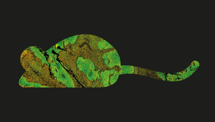
Glioblastoma accounts for over 60 percent of brain cancers, but survival five years beyond diagnosis is still rare. Raman and reflectance spectroscopies may allow us to distinguish healthy from diseased brain tissues with relative ease, improving our ability to detect and resect these tumors – and save patients.

After implanting murine glioma cells expressing enhanced green fluorescent protein into mice, Enrico Baria and colleagues used fluorescence microscopy to spot malignant tissue. Raman and reflectance spectroscopies aimed at the area of peak fluorescence intensity then informed a tissue classification algorithm, facilitating 97 percent identification accuracy when combining the two techniques.

The approach could provide a powerful tool for further animal studies – and could eventually complement preoperative MRI in humans.

Newsletters

Receive the latest analytical science news, personalities, education, and career development – weekly to your inbox.

Newsletter Signup Image

References

  1. E Baria et al., Neurophotonics, 7, 045010 (2020). DOI: 10.1117/1.NPh.7.4.045010.

About the Author(s)

Matt Hallam

I've always wanted a job that fosters creativity - even when I worked on the assembly line in a fish factory. Outside work, I satisfy this need by writing questionable fiction. The venture into science writing was an unexpected departure from this fiction, but I'm truly grateful for the opportunity to combine my creative side with my scientific mind as Editor of The Analytical Scientist.

More Articles by Matt Hallam

False

Advertisement

Recommended

False

False

The Analytical Scientist
Subscribe

About

  • About Us
  • Work at Conexiant Europe
  • Terms and Conditions
  • Privacy Policy
  • Advertise With Us
  • Contact Us

Copyright © 2025 Texere Publishing Limited (trading as Conexiant), with registered number 08113419 whose registered office is at Booths No. 1, Booths Park, Chelford Road, Knutsford, England, WA16 8GS.